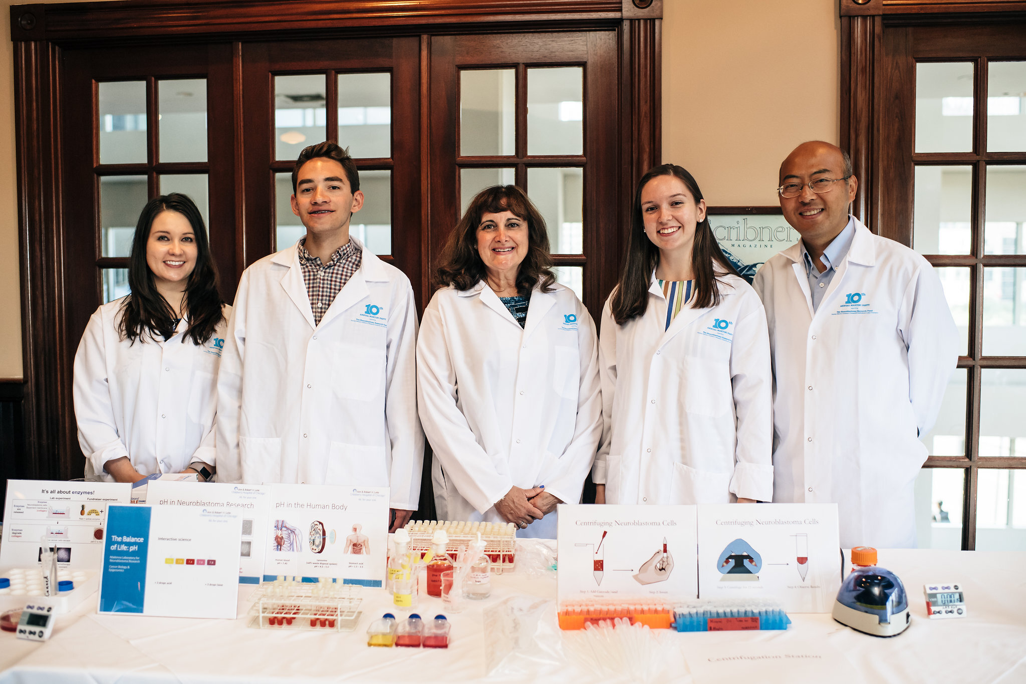
Neuroblastoma Pediatric Cancer Research
Dr. Mary Beth Madonna’s Laboratory
Dr. Madonna’s team has focused on studies to discover better ways to treat neuroblastoma and osteosarcoma.
The high mortality from NB is likely because many patients present with late stage, widely disseminated disease, carrying a poor prognosis of only 20-35% survival despite aggressive therapy. Osteosarcoma is the most common primary bone tumor of childhood accounting for 3-5% of childhood cancers with a 5-year survival less than 30% when metastatic or recurrent. Chemotherapy is the mainstay of treatment.
Although initial chemotherapy helps eliminate neuroblastoma and osteosarcoma in a substantial number of affected children, the majority of neuroblastoma and osteosarcoma patients eventually develop progressive disease that is resistant to further therapy. Novel therapies for advanced disease are urgently needed and our works intend to address this gap.
Neuroblastoma (NB) is the most common extracranial solid malignancy in childhood, accounting for nearly 10% of childhood cancers and 15% of cancer deaths in children.
Dr. Madonna's team has investigated new avenues to identify the basic biological processes that regulate adaptation of malignant tumor cells to stressful environments, such as those produced by anti-cancer drugs, and thereby permit their survival in the presence of agents that would normally kill them. Understanding these processes will help cancer researchers develop novel chemotherapeutic strategies.
Dr. Madonna and her team have made considerable progress pursuing several different avenues of research, highlighted below, to reach the ultimate goal of finding new ways to screen for and effectively treat neuroblastoma and osteosarcoma.
1. Expression of checkpoint inhibitors in neuroblastoma and osteosarcoma and potential targets for treatment
Inhibition of immunomodulatory checkpoint molecules, molecules that normally prevent the immune system from inappropriately reacting with normal cells, has been a successful, novel treatment for chemo-resistant and metastatic cancers in adults. Cancer can hijack these regulatory pathways to prevent immune recognition and destruction of cancer cells. Although many checkpoint molecule receptors exist, inhibition of several including CTLA-4, PD-1, and PD-L1 has shown promise treating advanced melanoma, non-small cell lung cancer, and multiple other cancers common in adults. The use in pediatric solid tumors is still in its infancy. Dr. Madonna’s laboratory is uniquely situated to examine surplus tumor materials from patients due to RUSH’s strong clinical practices and banking of bio-specimens. Profiling immunomodulatory molecules is required to understand the immune system’s response to cancer, which will help illuminate immunotherapy’s role as a promising therapeutic approach for osteosarcoma and neuroblastoma.
Current findings and future work:
We have demonstrated that the checkpoint molecule PD-L1 is expressed in both wildtype and drug-resistant neuroblastoma cells from three different cell lines. Moreover, it is expressed in a drug-resistant osteosarcoma cell line but not in the wildtype cell line. This makes PD-L1 an excellent potential target for treatment in both cancers. This research will be presented at the Academic Surgical Congress in February, 2021. Since our neuroblastoma and osteosarcoma cell lines both expressed PD-L1 to varying degrees, we are currently testing whether inhibition of the PDL1 receptor on cancer cells in vitro works to improve chemotherapy treatment and reduce cancer invasiveness. We are also in the process of testing each of these cell lines for 15 other checkpoint molecules that could be targets for drug treatment too.
In this project, we are studying osteosarcoma and neuroblastoma together, since they are common pediatric solid tumors with limited treatment options for metastatic or recurrent disease, and typically have high T-cell infiltrates— a common metric used to determine responsiveness to checkpoint inhibition.
2. Investigation of HDAC-6 regulation of HIF-1α and the role it plays in mediating both drug resistance and invasiveness in osteosarcoma and neuroblastoma
Osteosarcoma and Neuroblastoma are common childhood cancer with a poor prognosis when recurrent or metastatic. Prior studies suggest that multi-drug resistant tumors are more metastatic relative to their parental wild-type cells but the mechanism remains unknown. There is ample evidence that solid tumors frequently encounter hypoxic stress. Rapidly proliferating cancer cells may outgrow their vascular network, limiting O2 diffusion within the tumor. Importantly, hypoxia has been associated with drug resistance and reduced sensitivity to radiation therapy. Cellular response to hypoxia is controlled in part by hypoxia inducible factor-1α (HIF-1α). HIF-1α is recognized as a key modulator of the transcriptional response to hypoxic stress.
Neuroblastoma often exhibits high levels of HIF-1α accumulation. Dr. Madonna’s team has found that hypoxia indeed plays a key role in the development of drug resistance, further research in this area may lead to new therapies that are able to overcome drug resistance of doxorubicin in clinical settings. Many studies show that histone deacetylases (HDACs), which are epigenetic regulators of gene transcription play a role in tumorigenesis and may regulate activity of HIF-1α which controls gene expression associated with drug resistance and metastasis. Currently there are histone deacetylase inhibitors in phase I and II drug trials and could be used as chemotherapy. We hypothesized that this same mechanism functions as a link between chemoresistance and invasiveness in osteosarcoma.
Current findings and future work:
Via various laboratory techniques we have demonstrated that drug resistant osteosarcoma and neuroblastoma cells are more invasive than their parent cells. That is to say, these drug resistance cells behave more like metastatic cancer. The drug resistant cell lines not only demonstrate upregulation of HDACs and HIF-1α, but we have demonstrated that these pathways are linked. Ultimately, HDAC inhibitors reduce invasiveness of drug resistant cells and may provide an adjuvant treatment. This work was presented at the American Pediatric Surgical Association’s Annual Meeting and is currently submitted for publication.
3. Targeting the Notch signaling pathway to overcome drug resistance in neuroblastoma cells
Recently, emerging evidence suggests that the Notch signaling pathway is one of the most important signaling pathways in drug-resistant tumor cells. Moreover, down-regulation of the Notch pathway could induce drug sensitivity, leading to increased inhibition of cancer cell growth, invasion, and metastasis. Therefore, the targeting of Notch pathways may be a novel therapeutic approach for treatment of cancer by overcoming drug resistance. This can lead to the elimination of cancer stem cells (CSCs) or epithelial to mesenchymal transition (EMT) type cells which are typically drugresistant, and are thought to be the root cause of tumor recurrence. To date, there have been no studies examining the effects of γ-secretase inhibitors (GSI) on the anti-cancer effects in drug resistant human neuroblastoma tumor cells.
Current findings and future work:
Dr. Madonna’s lab found N-[N-(3,5-Difluorophenacetyl)-L-alanyl]-S-phenylglycine t-butyl ester (DAPT), a PanNotch and γ-secretase inhibitor (GSI), significantly potentiated doxorubicin-induced cytotoxicity in SK-N-SH and SK-NBE(2)C drug resistant neuroblastoma tumor cells. They further demonstrated that DAPT synergized with doxorubicin and significantly reduced the growth of DoxR neuroblatoma cells possibly via inducing senescence, these data were further confirmed by senescence SA-β-Gal staining. Dr. Madonna’s lab also tested another GSI, COMP E, and has found out that COMP E worked the same as DAPT to overcome drug resistance in neuroblastoma tumor cells. These findings further supported that inhibiting Notch signaling pathway by GSI could be a promising approach for dealing with drug resistant neuroblastoma and may have a great clinical significance.
To unveil the underlying mechanism of the GSI’s effects, Dr. Madonna’s lab compared the proteomic profile of DoxR NB cells pretreated by DAPT with that from cells pretreated by both DAPT and doxorubicin. As reported previously, through the use of a two-dimensional-differential-in-gel-electrophoresis (2D-DIGE) approach applied for the first time to 3 the screening of proteomic expression in drug resistant neuroblastoma tumor cells, they identified 9 unique proteins in 13 spots differentially expressed and associated with inactivation of Notch signaling pathway.
These unique proteins could be potentially considered as useful diagnostic markers and therapeutic candidates for drug resistant neuroblastoma tumor treatment. This finding further demonstrated that Notch signaling might play a pivotal role in regulating different pathways which contribute to the development of drug resistance in NB cells. We anticipate that this work will pave the way in the near future to evaluate the efficacy of targeting Notch signaling pathway in enhancing the response of NB to chemotherapy treatment.
4. Detection of a model system using gold nanoparticles
An avenue to fighting cancer in its many forms is to catch it early and start treatment before tumors get too large or metastasize to other parts of the body. Mostly the cancer is caught too late and the mortality rate increases. One way that researchers have attempted to decrease the mortality rate is by finding ways to detect the cancer and treat sooner and more effectively. One such route that has been looked at for this early detection is through the use of nanoparticles to bind to specific biomarkers within the body. Instead of biomarkers, this project uses a model system where gold nanoparticles coated with biotin are put in solution with streptavidin. Due to the strong binding affinity streptavidin has for biotin, the two molecules bind strongly and bring the nanoparticles close together causing a change in color of the solution.
Current findings and future work:
Past work on this project has shown an observable change in absorbance with addition of streptavidin to the solution. Dr. Madonna’s lab plans to replicate these results and expand upon them by observing how variations in conditions such as buffer solution, pH, and temperature affect resistance in NB cancer cell lines. These variations in conditions serve to better replicate physiological conditions to ensure that the nanoparticles would still be usable in the body.
Dr. Madonna’s research team is also planning to use electron microscopy to image the nanoparticles to show the nanoparticles coagulated together. Preliminary runs have shown that there is little to no interaction between the gold nanoparticles and the biotinylated gold nanoparticles when in the deionized water and phosphate buffer solutions. This ensures that the changes in absorption are in fact due to the addition of streptavidin.


